Dive into the Diagnostic Breakthroughs with ZEISS CLARUS
Explore Case Studies by Leading Experts
Explore the insightful case studies of Dr. Aditya Kelkar, Dr. Vishal Agarwal, and Dr. Avnindra Gupta that showcase the color, clarity and precision of ZEISS CLARUS, elevating diagnostic excellence. Uncover diagnostic inights, and treatment plans that elevate ophthalmic practice and improve patient outcomes.
Unlock Complex Eye Conditions and Enhance Your Ophthalmic Practice
Explore Clinical Cases by Dr. Aditya Kelkar
Clarus clinical case image
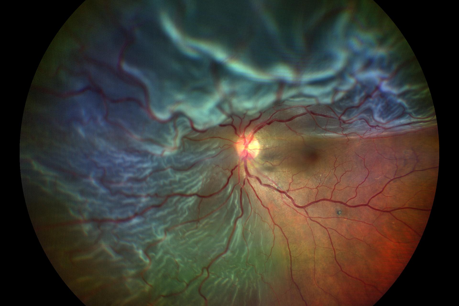
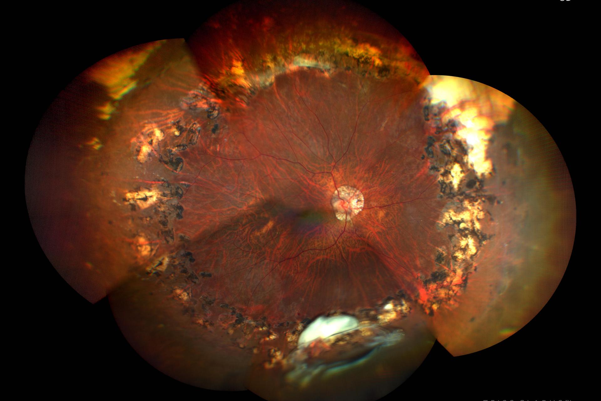
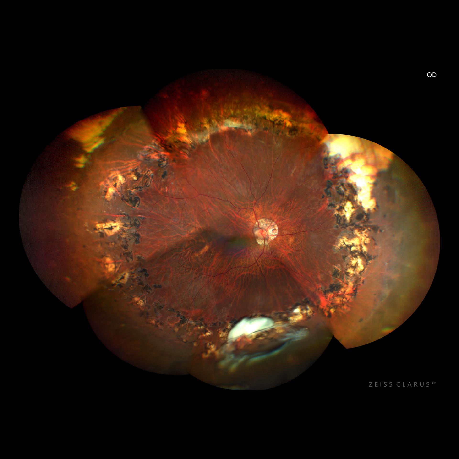
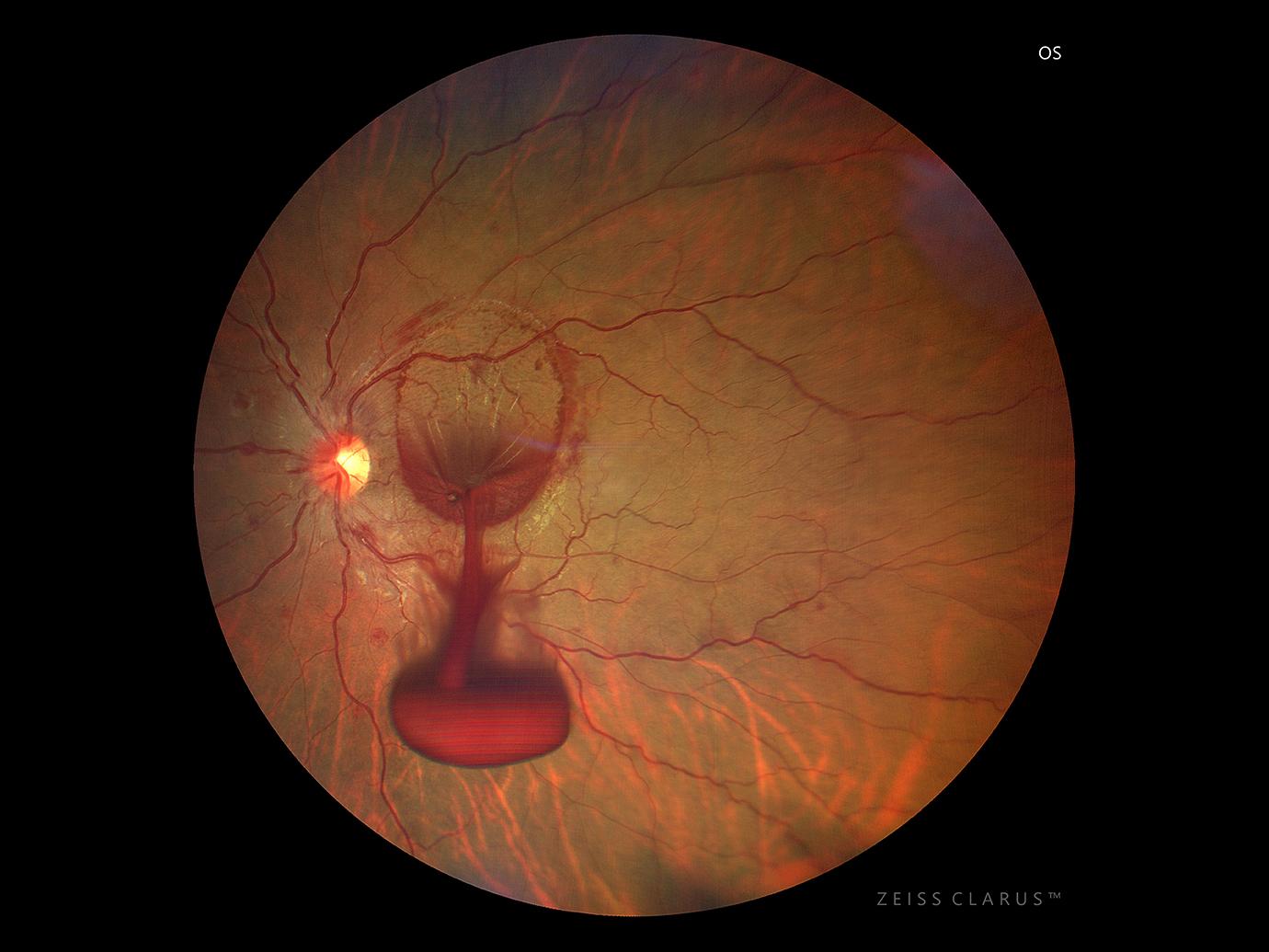
Rhegmatogenous Retinal Detachment
A 70-year-old patient came in OPD with a complaint of blurring of vision with positive scotoma in inferior gaze for the past 4 days. Color fundus photograph of the left eye showed Rhegmatogenous Retinal Detachment with the Macula Off.
Please click the arrow to view next case study.
Clarus clinical case image
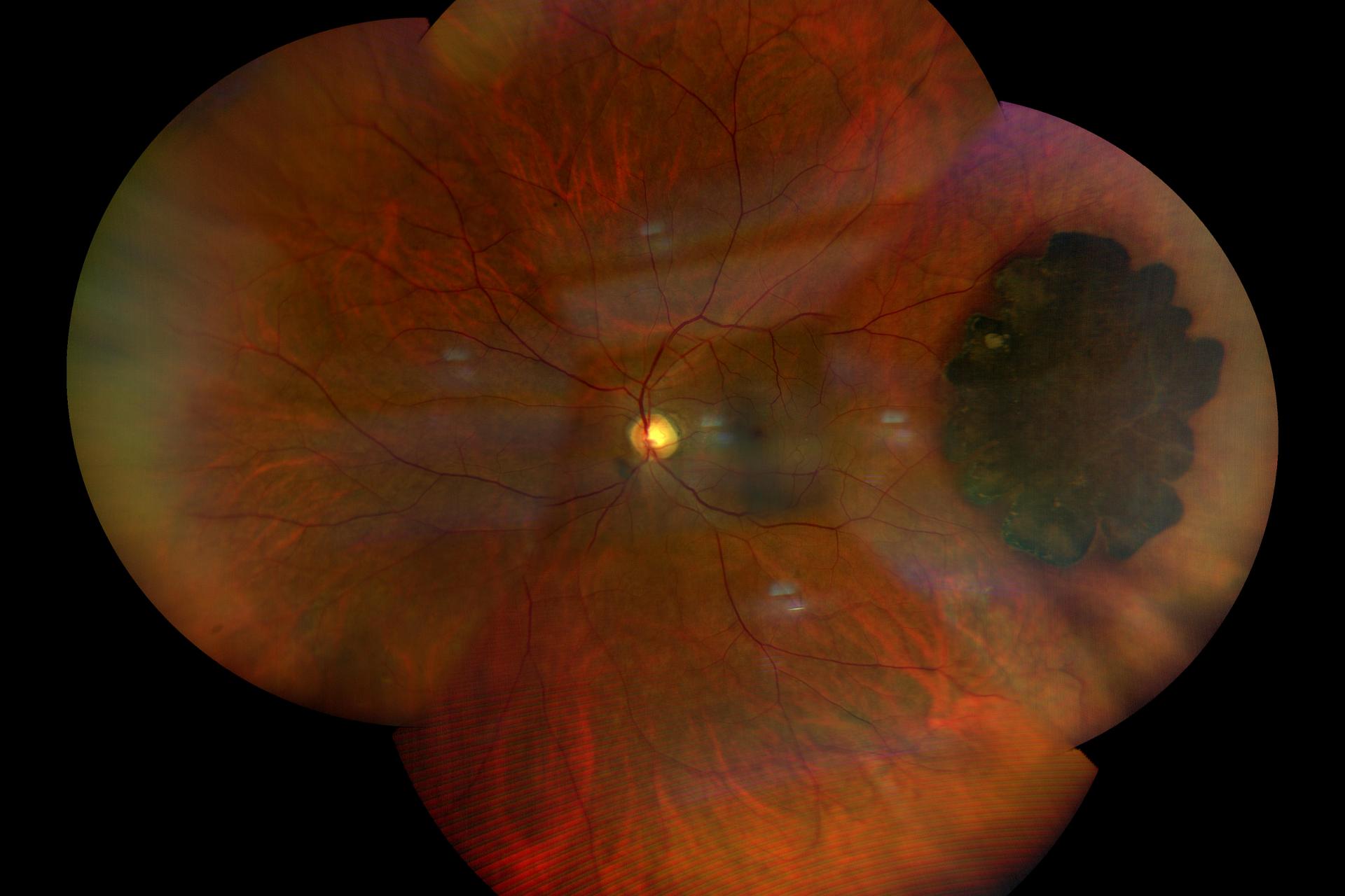
Choroidal Nevus
A 47-year-old male came in OPD with a complaint of floaters in the left eye for the past 1 year. Color fundus photograph of the left eye showed large Choroidal Nevus.
Clarus clinical case image
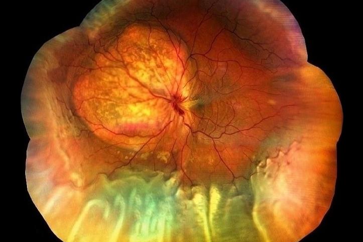
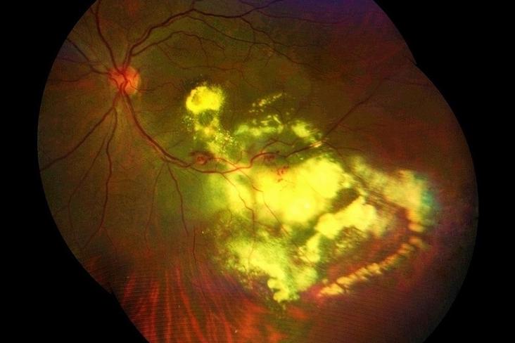
Choroidal Melanoma with Exudative Retinal Detachment
A 45-year-old male came in OPD with a complaint of progressively diminishing vision in his left eye over the past few months, which is worsening. Color fundus photograph of the left eye showed Choroidal Melanoma with Exudative Retinal Detachment.
Clarus clinical case image
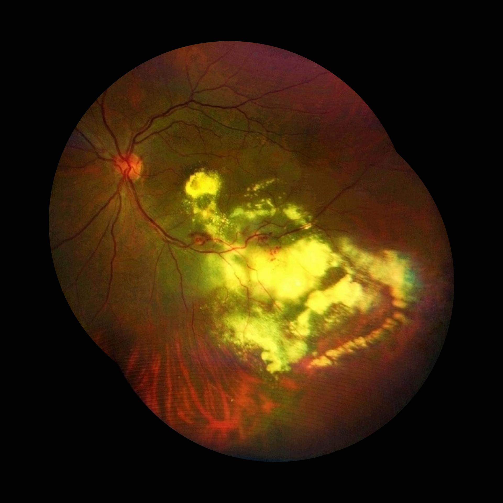
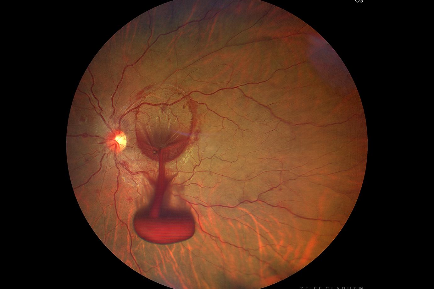
Leber's Miliary Aneurysm
A 53-year-old female came in OPD with a complaint of floaters and diminishing vision in her left eye for the past one month. Color fundus photograph of the left eye revealed Leber’s Miliary Aneurysm.
Leber’s Miliary Aneurysm is a vascular anomaly characterized by vascular telangiectatic lesions and Retina Exudation.
The ZEISS CLARUS has transformed my ophthalmology practice. Its ultra-widefield imaging technology provides a stunning panoramic view of the retina in true color, revealing ocular conditions that traditional methods often miss. The exceptional clarity and detail of the peripheral retina images are game-changers, allowing me to identify serious retinal diseases at an early stage. With the ZEISS CLARUS, I can diagnose and monitor these conditions with unprecedented confidence, ensuring my patients receive the highest standard of care.
Clinical Cases by Dr. Avnindra Gupta
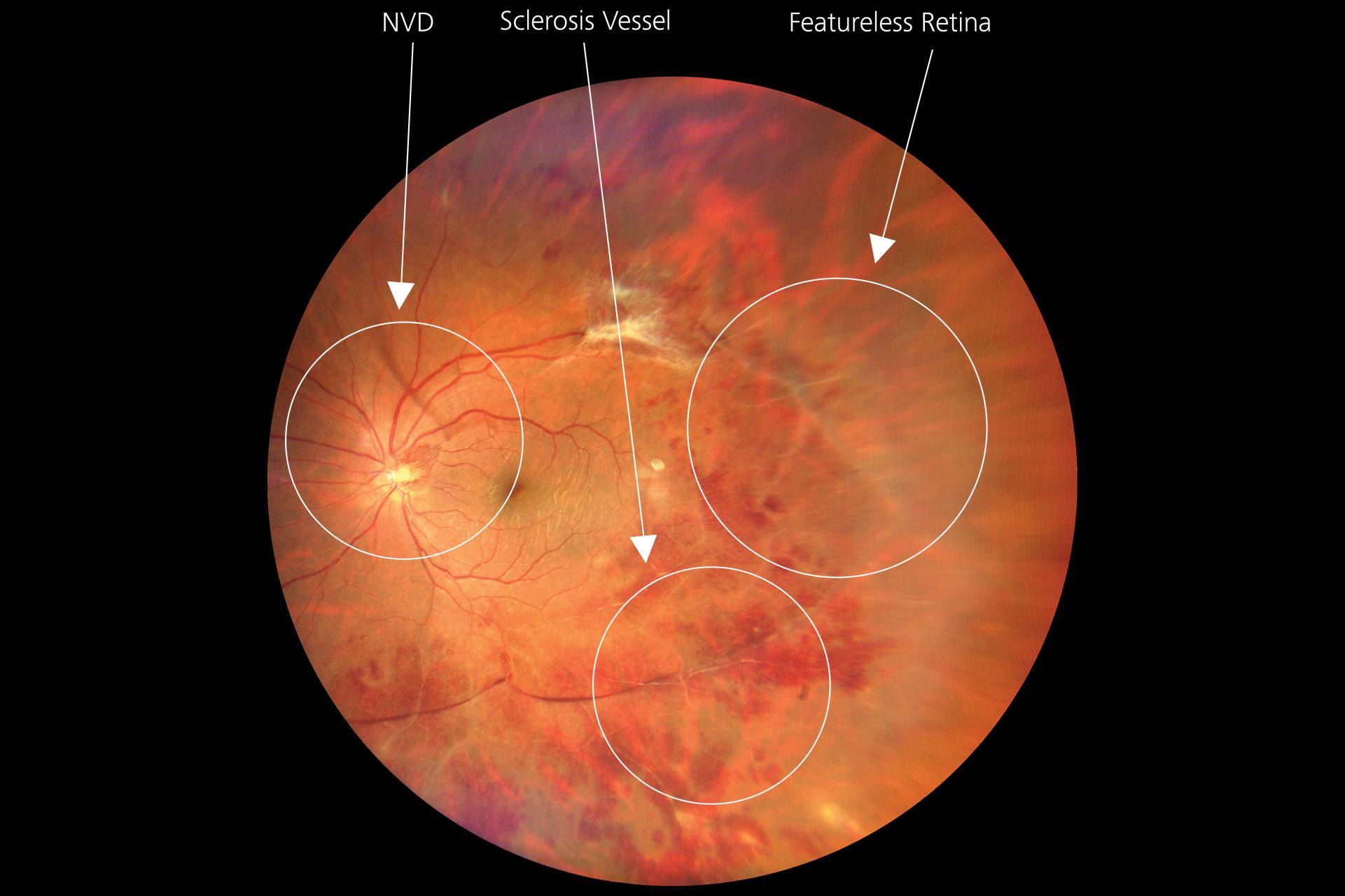
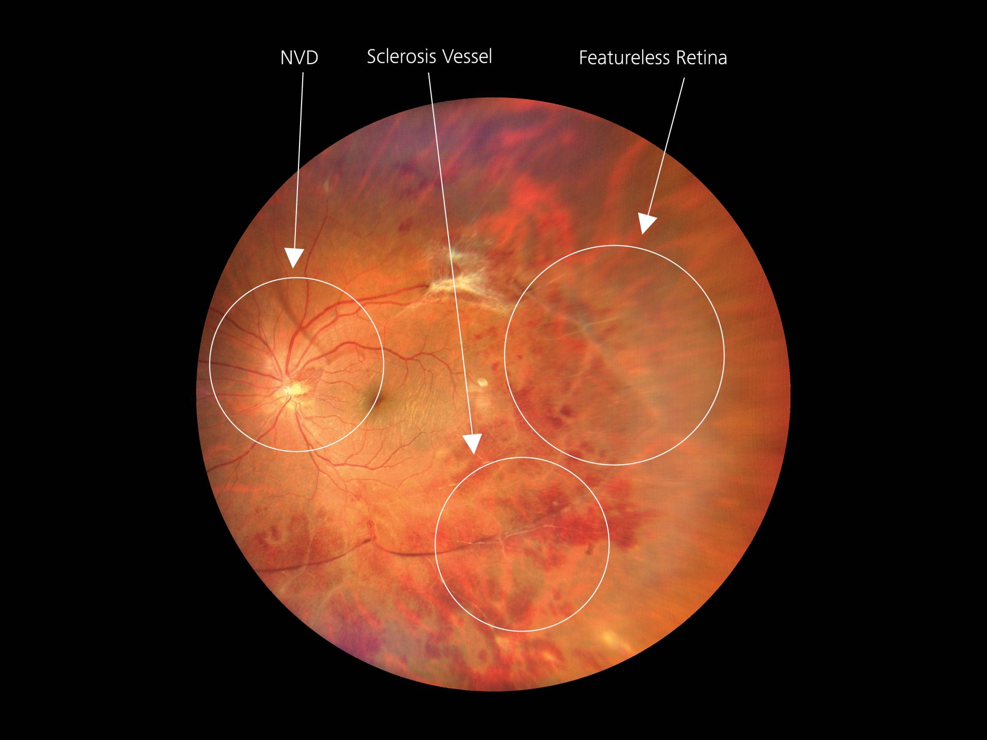
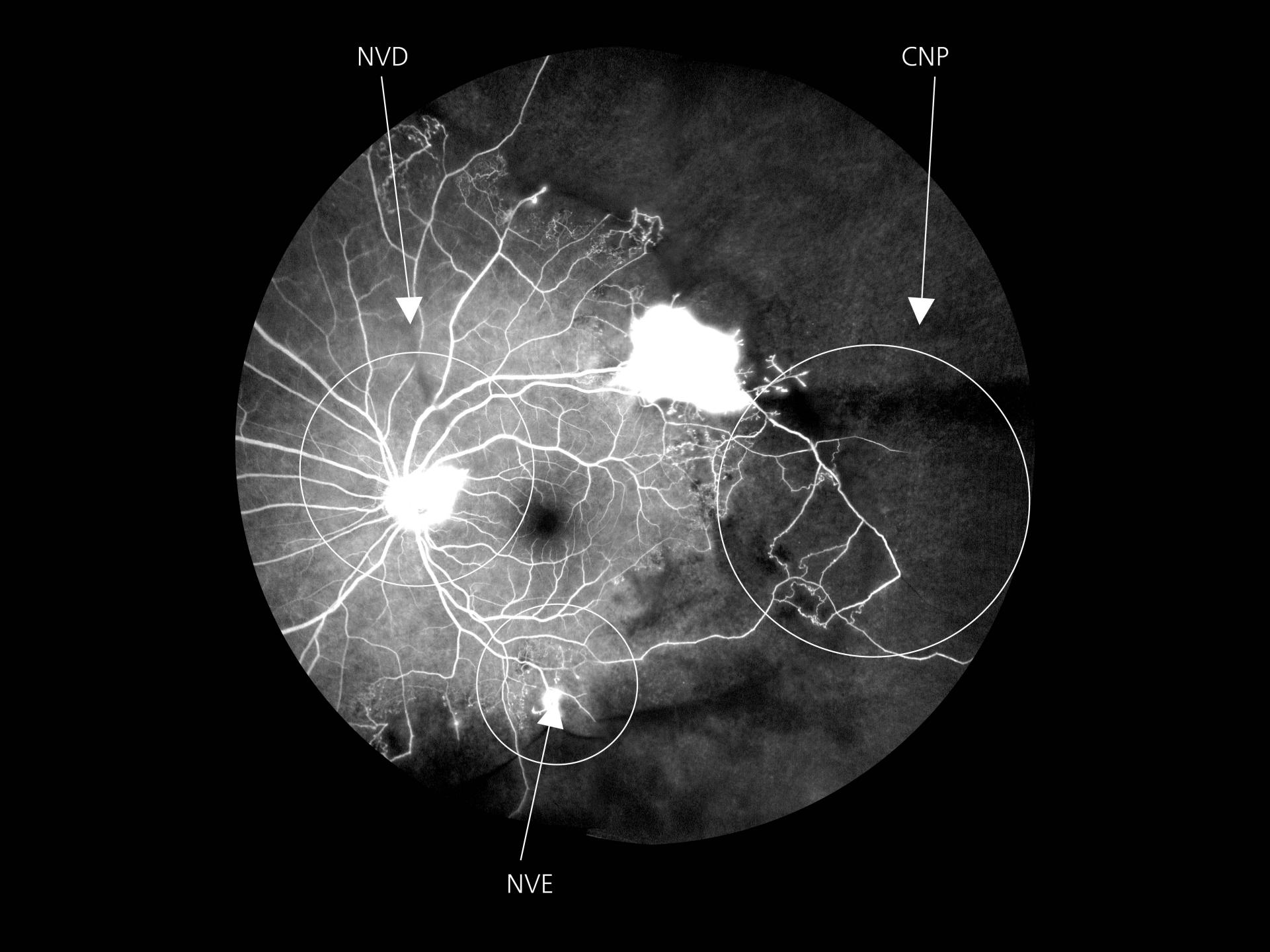
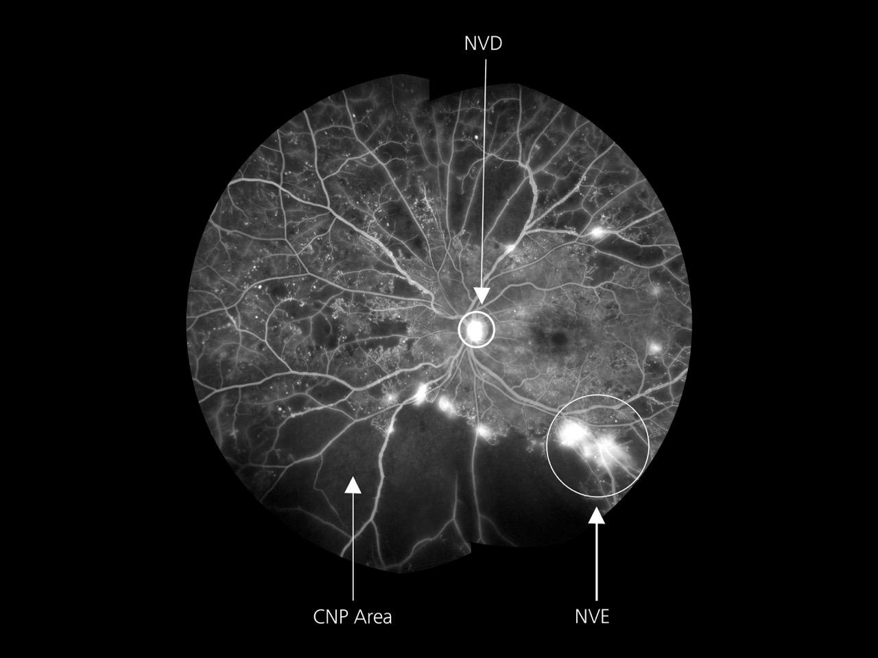
Eale’s Disease
A 40-year-old male patient presented with defective vision and floaters in the left eye.
UWF true color images indicate a tuft of new blood vessels near the disc called neovascularization disc (NVD) with multiple hemorrhages all over the fundus and sclerosed blood vessel at the superior and inferior arcade. The Gaze Point feature helped in tracing the sclerosed blood vessels leading to CNP areas outside the arcade in the temporal quadrant.
Please click the arrow to view the next case study & slider to view next image.
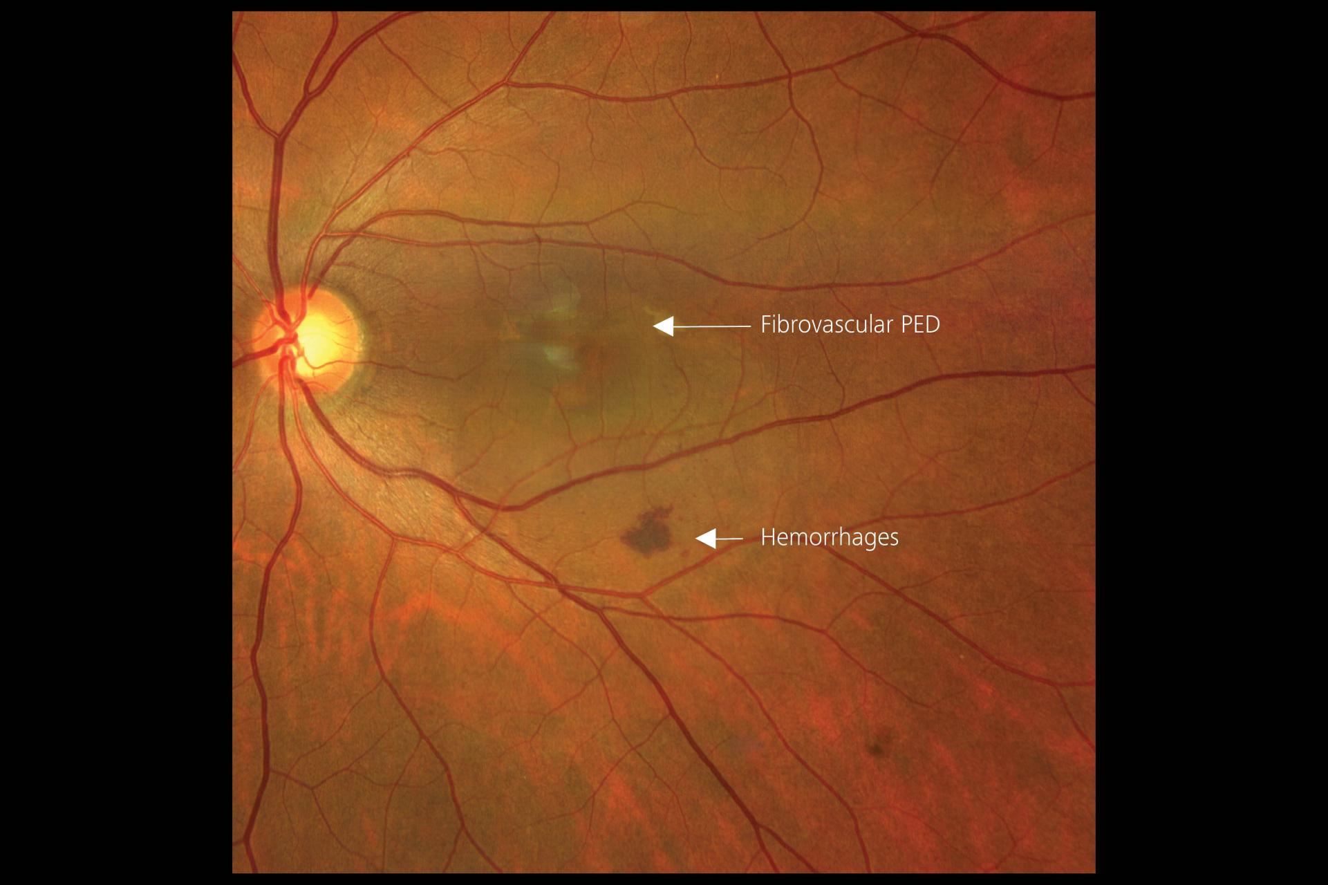
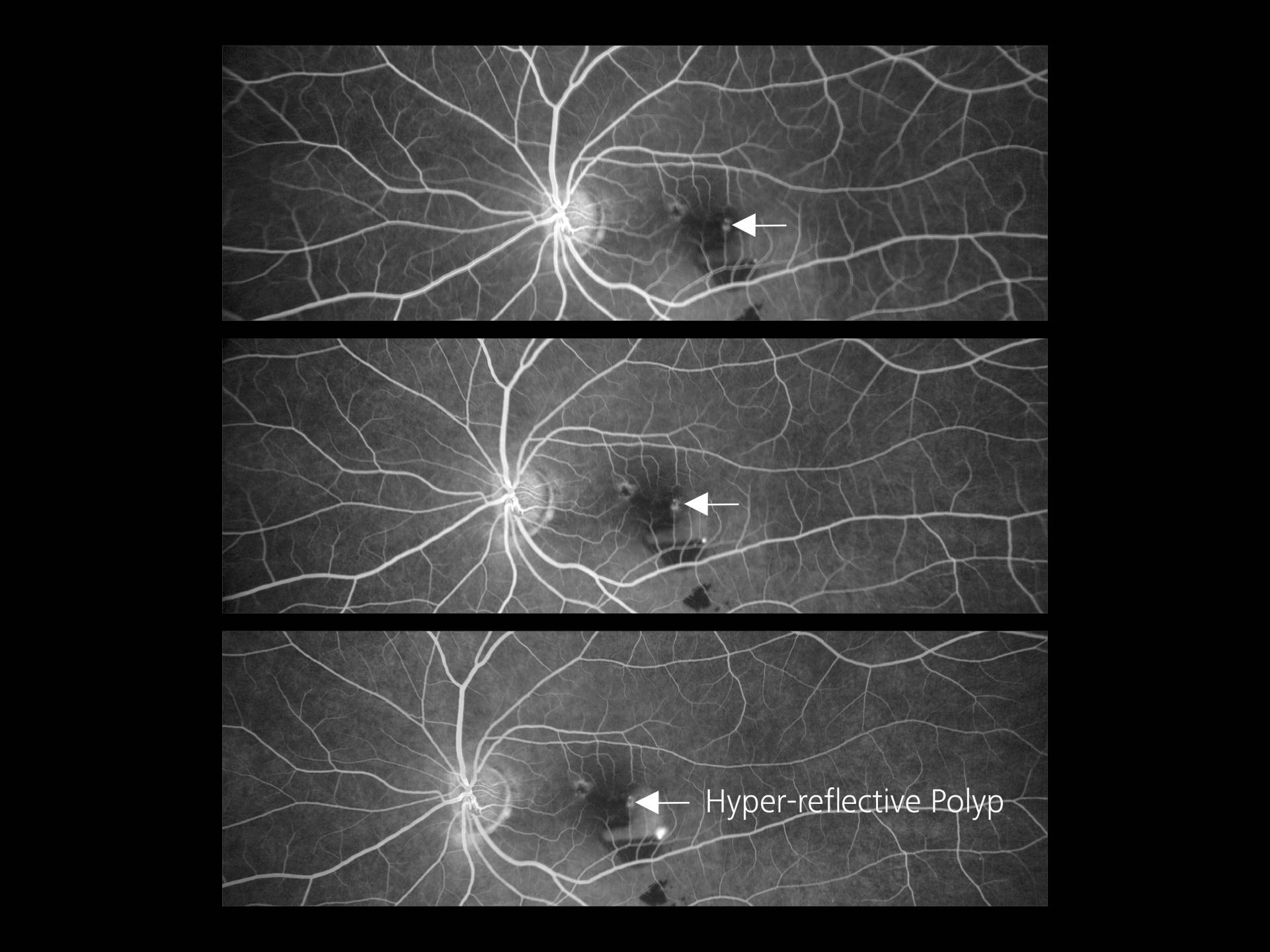
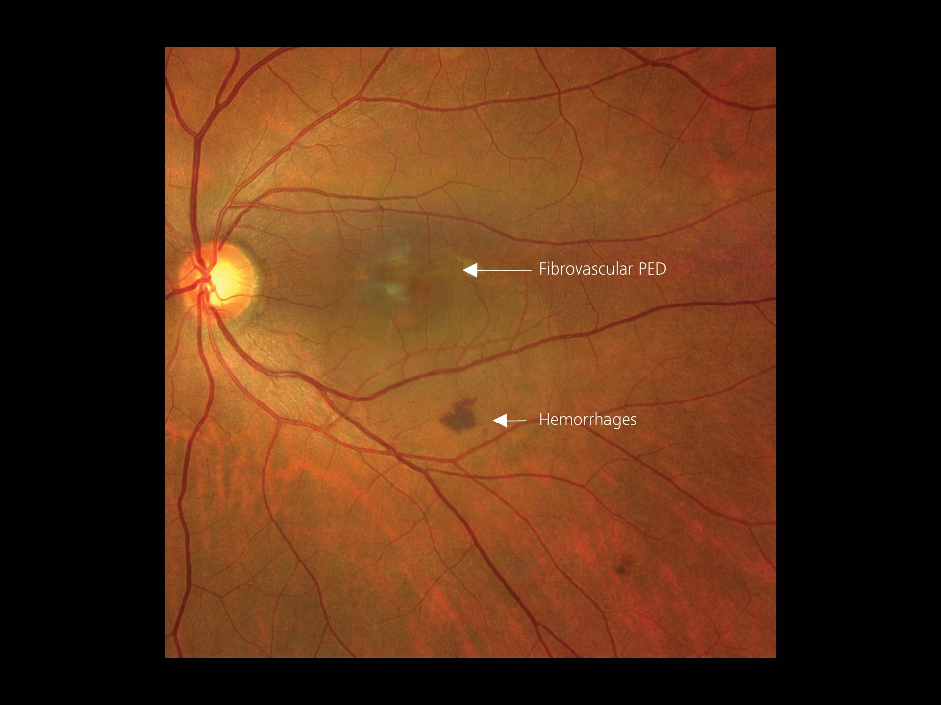
ARMD with PCV (Polypoidal Choroidal Vasculopathy)
A 54-year-old male patient presented with defective vision in the right eye and no associated systemic illness was reported.
A single widefield (133°) true color image on the ZEISS CLARUS 700 clearly indicates macular edema with hemorrhage. The Precision Focus feature of ZEISS CLARUS helped to attain high-resolution images even at high magnification showing even the smallest hemorrhage spots. The True Color – Fundus imaging clearly differentiated PED part from the rest of the healthy Retina. Upon further investigation, the Venous Phase of FFA (Fundus Fluorescence Angiography) showed a hyperfluorescent spot that indicates polyp within the fibrovascular PED. (Polyp) is generally seen with the ICGA but the high resolution of ZEISS CLARUS shows POLYPS in the FFA.
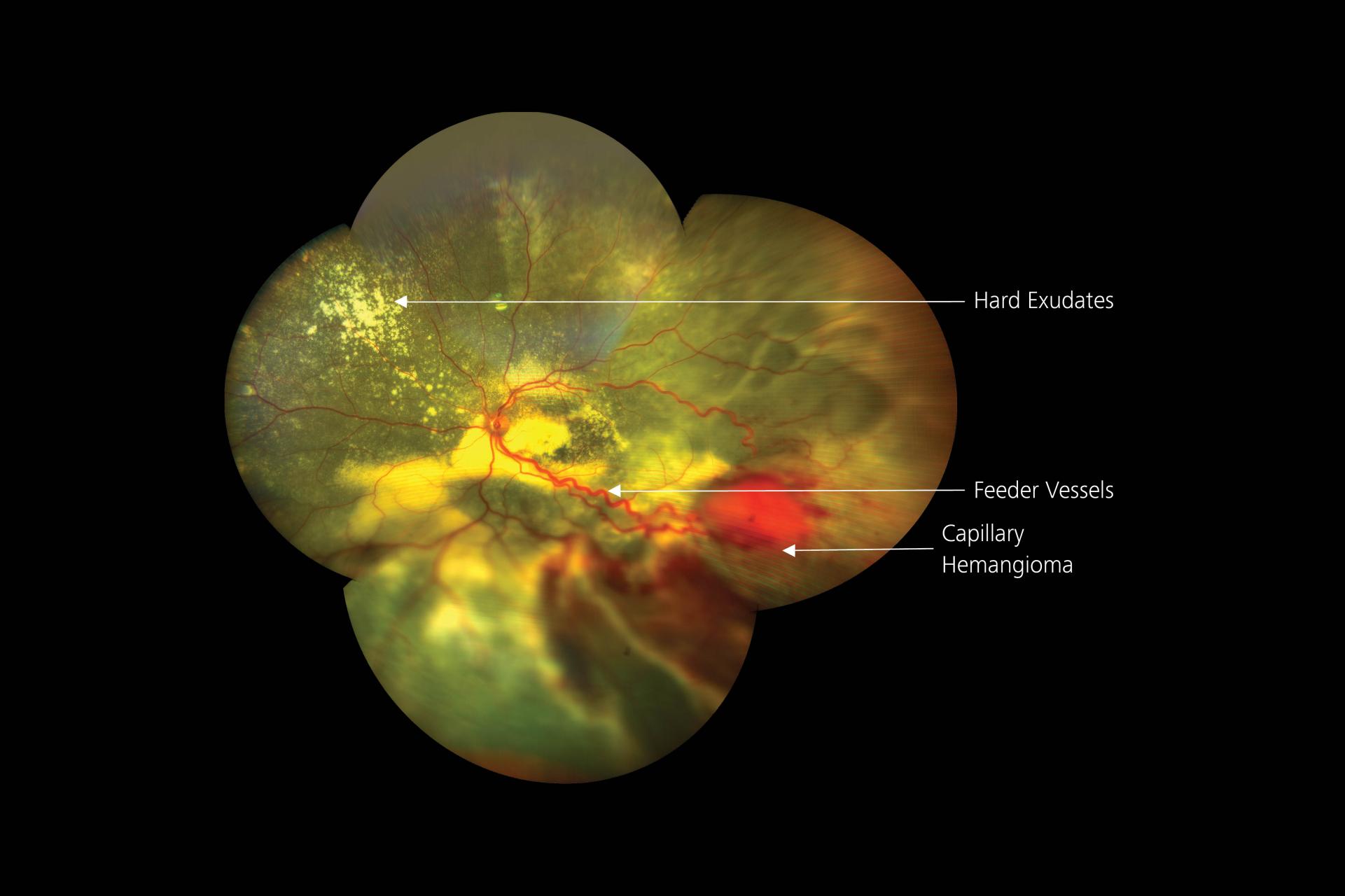
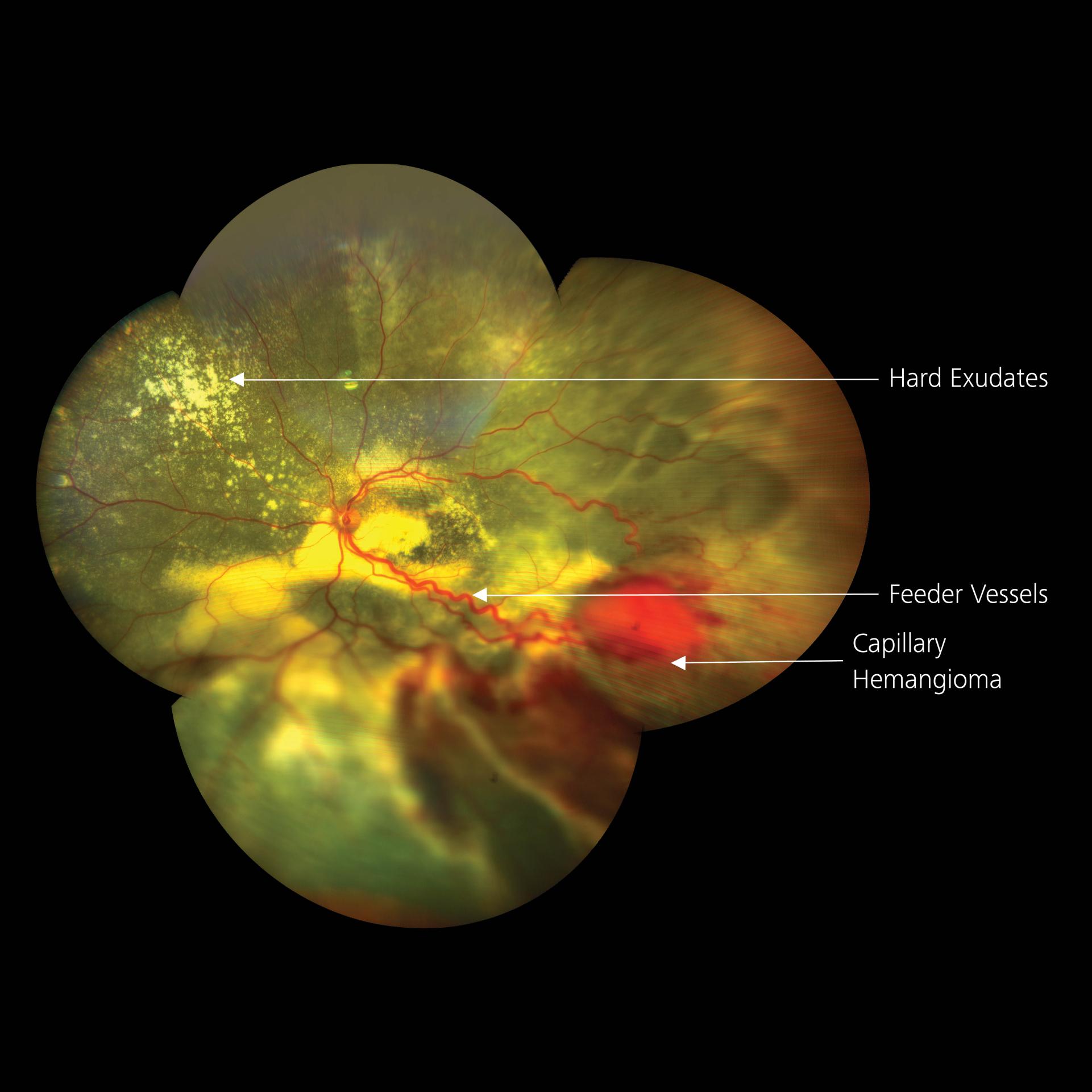
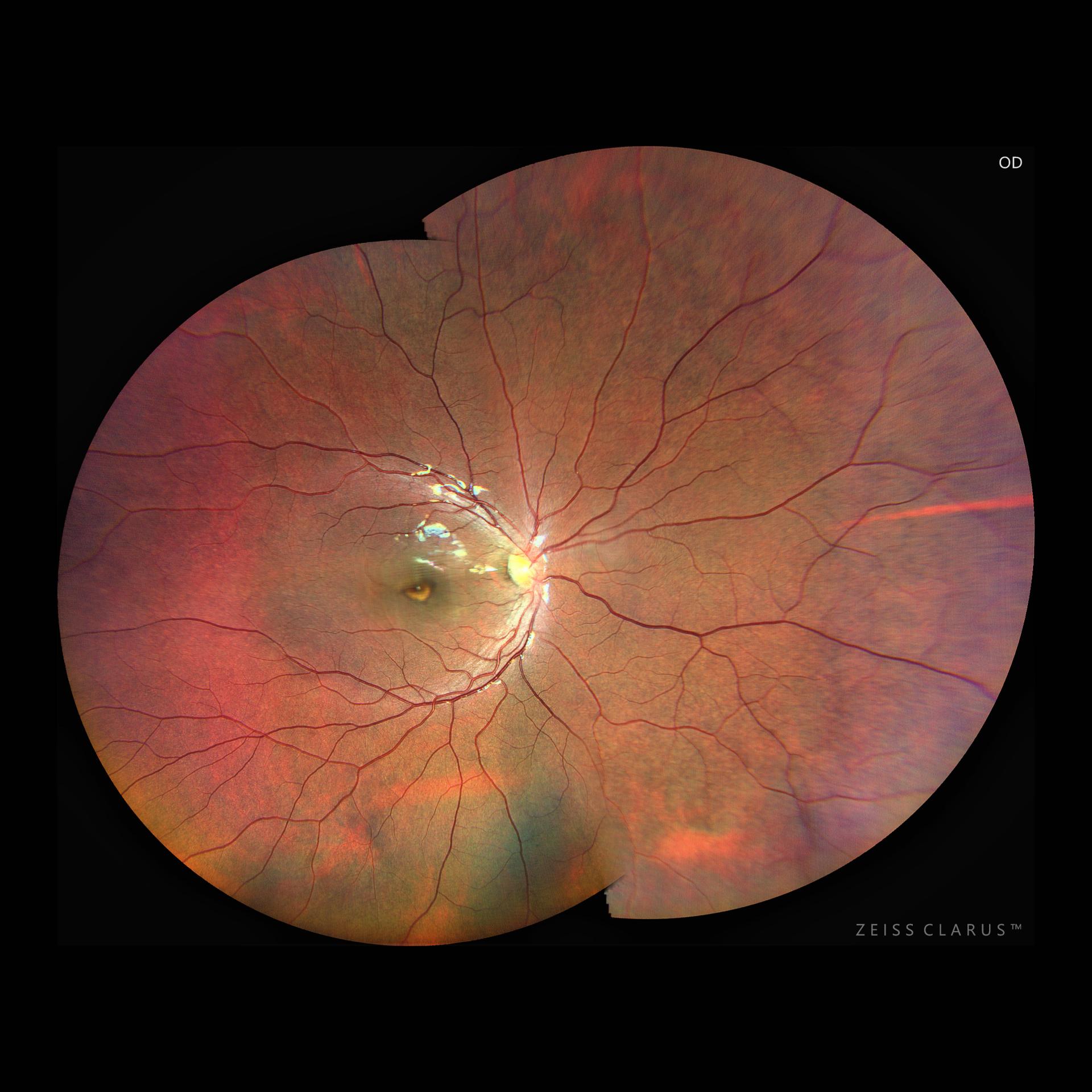
Capillary Hemangioma
An 8-year-old male presented with diminished vision in his right eye and eye divergence. A systemic examination, including an MRI, revealed the presence of a medullary hemangioblastoma. True Color fundus images on ZEISS CLARUS clearly indicate the Capillary Hemangioma, present in Infero-temporal quadrant (ITQ) with a large feeder vessel.
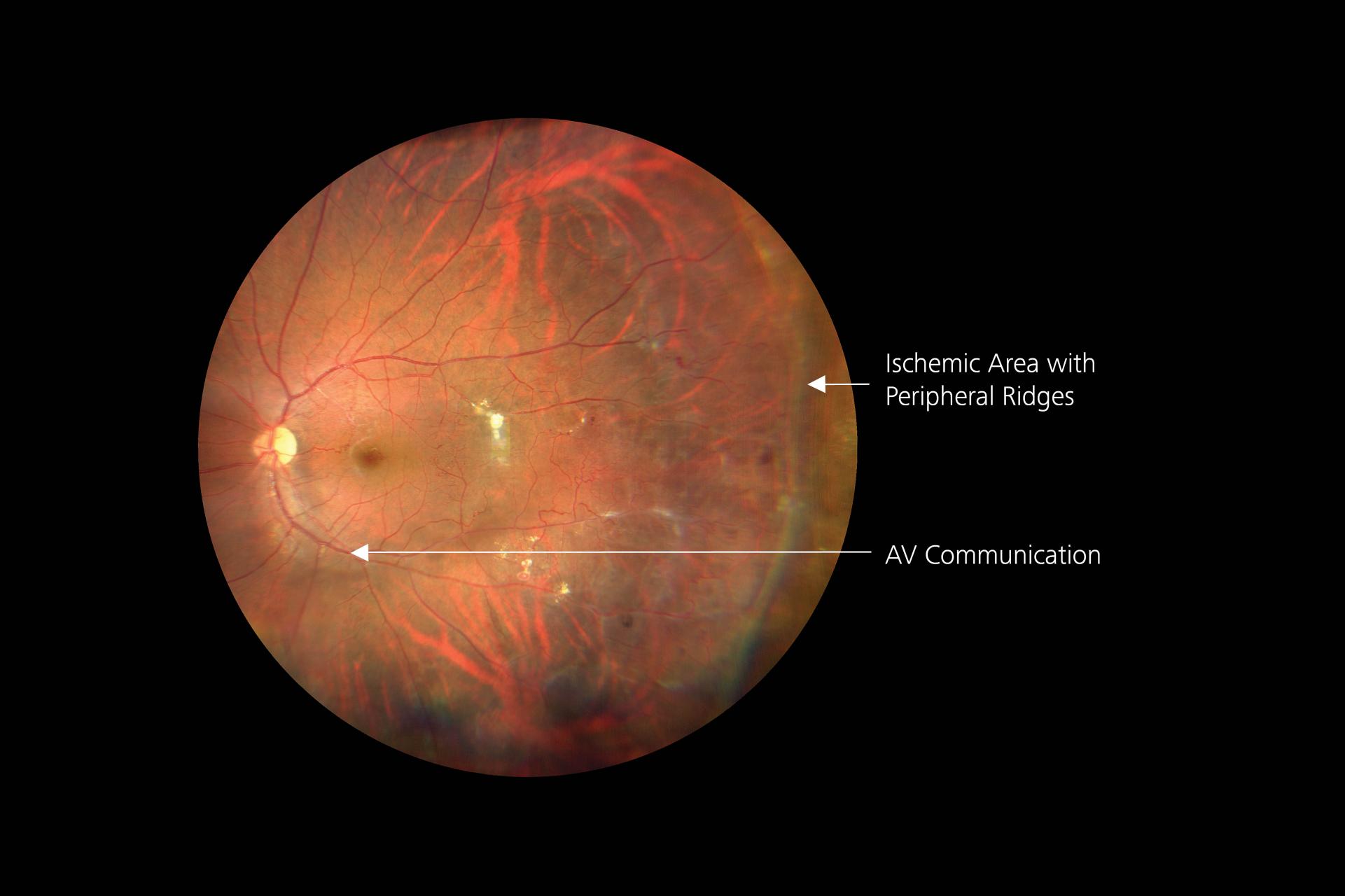
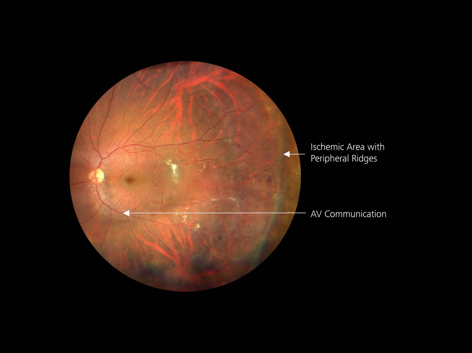
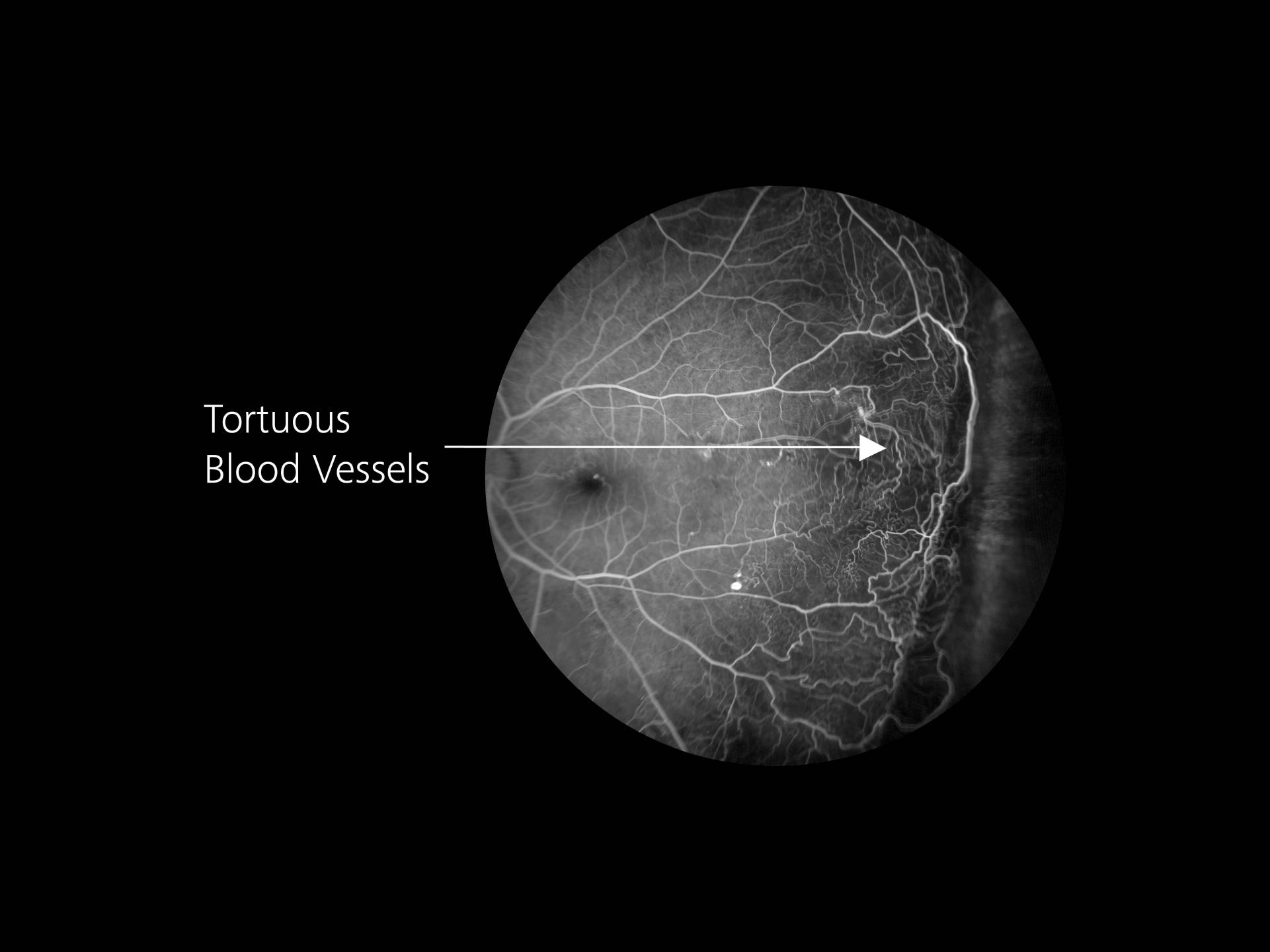
Familial Exudative Vitreoretinopathy (FEVR)
A 32-year-old male patient presented with complaints of floaters in both eyes. His systemic history was unremarkable, and no associated illnesses were noted.
Widefield images were taken of the central retina as well as the peripheral retina. Tortuous blood vessels were noted in all quadrants, and a demarcation line called RIDGE separated the vascular retina from the ischemic or avascular retina. The Auto-bright feature helped to maintain effective brightness throughout the angiogram, from early to late frames, for optimum visualization of tortuous vessels with AV communication in the peripheral part of the retina. Peripheral areas could be seen effortlessly and focused with the help of the Precision Focus feature. It helped to show capillary non-perfusion with ridges providing greater details.
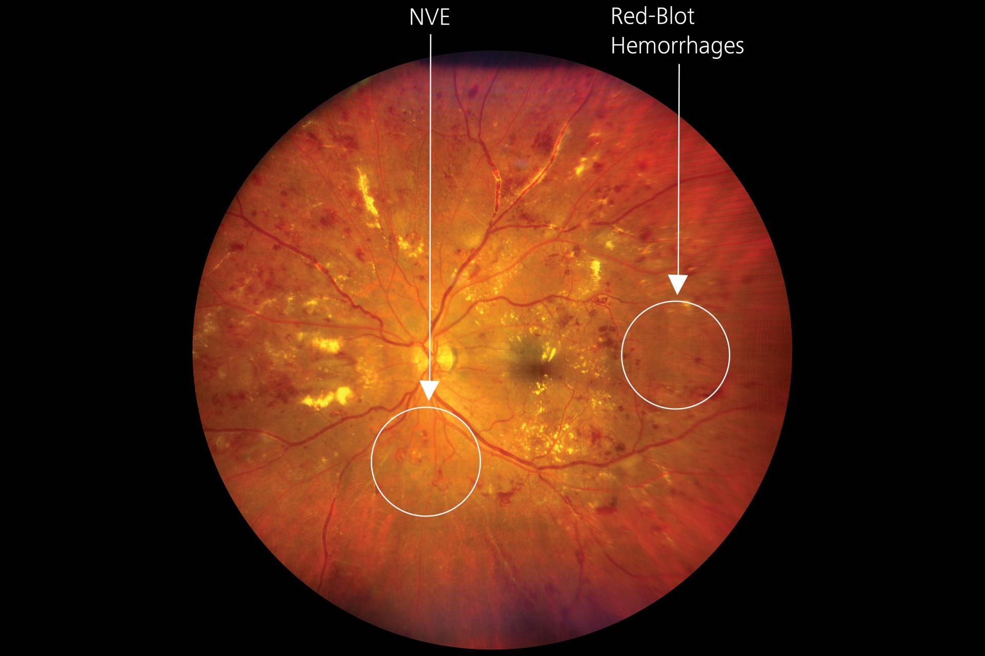
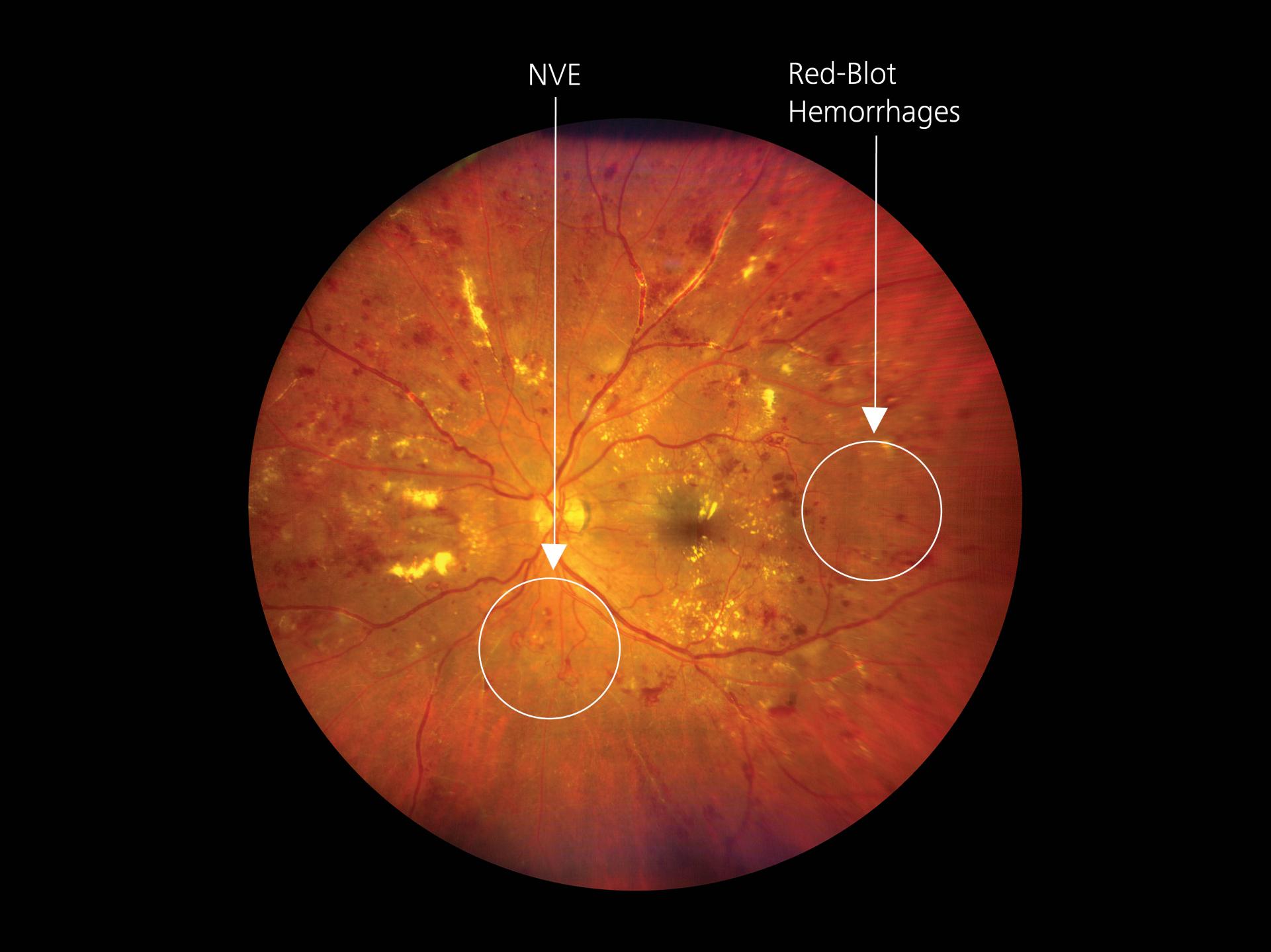
Proliferative Diabetic Retinopathy with Macular Edema
A 45-year-old male patient was seen with the complaint of a vision defect in both eyes. His systemic history revealed the presence of Diabetes Mellitus for 8 years. A single Widefield image (133 degrees) of ZEISS CLARUS was sufficient to cover the ETDRS 7 Field (an area essential for Diabetic Retinopathy screening and classification). True Color high-resolution images showed multiple Dot and Blot Hemorrhages (DBHs) Cotton Wool Spots (CWS), hard exudates and neovascularization. Areas of non-perfused retina (CNP) could be clearly seen in the peripheral part of the retina, showing neovascularization and CNP areas of the peripheral retina while maintaining resolution and sharpness.
Delving into the world of Retina and Fundus imaging with ZEISS CLARUS 700 has been truly enlightening. Crafted by the renowned optics manufacturer ZEISS, the technology delivers an exceptional one-shot, full high-resolution, and true-colored view of the retina. My experience with ZEISS CLARUS 700 has provided invaluable insights, setting a new standard in ultra-widefield retinal imaging.
Clinical Cases by Dr. Vishal Agarwal
Clarus clinical case image
Move the slider to view next image
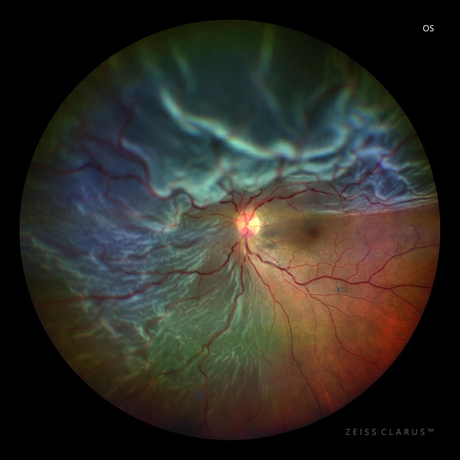
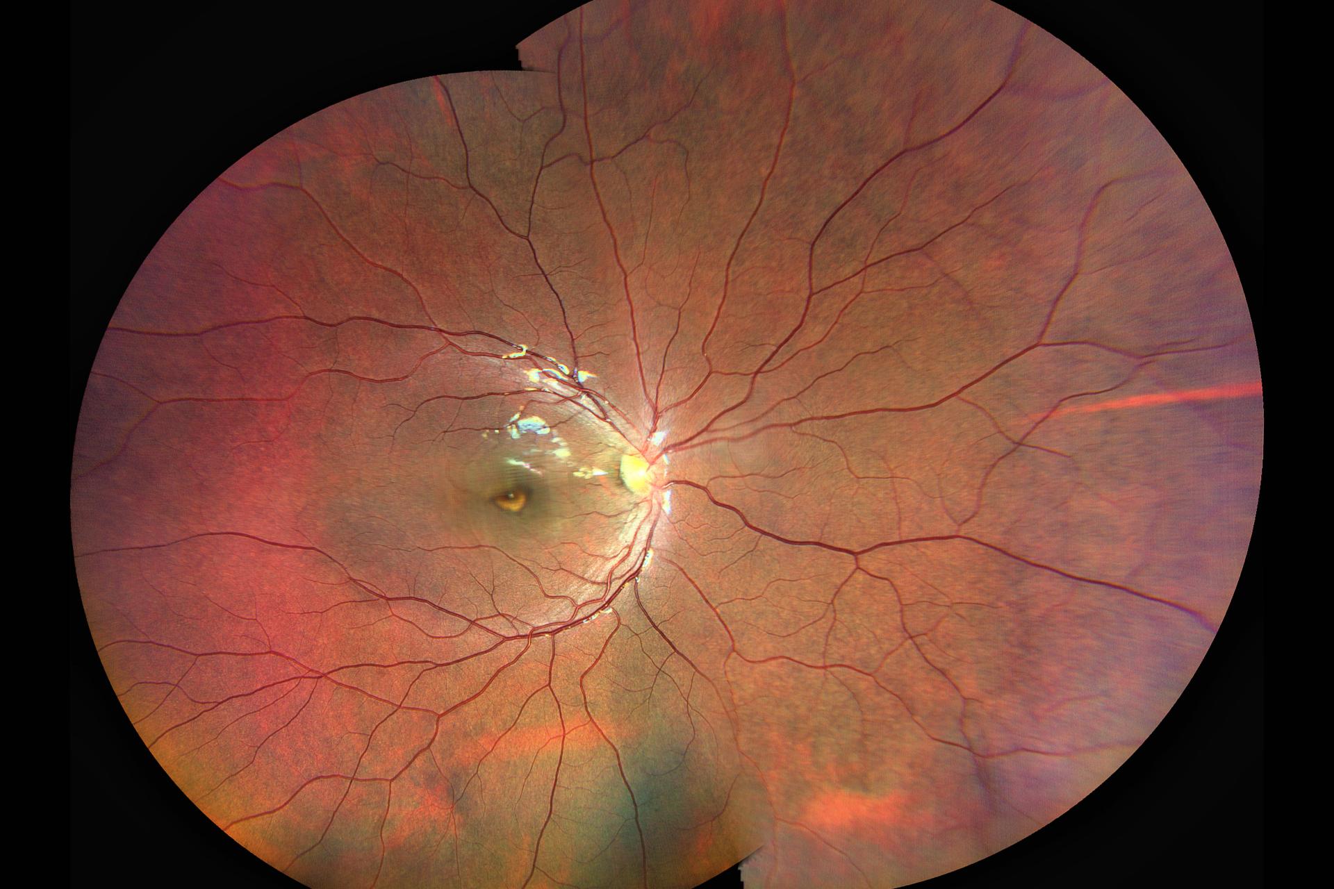
Case of Bull’s Eye Maculopathy in Cone Dystrophy
Retinal analysis of a 12-year-old boy with complaint of diminishing vision and nystagmus. True color ultra-widefield fundus imaging helped in detecting Bull’s Eye Maculopathy suggesting Cone Dystrophy. On widefield imaging it appeared like an eye on macula due to nystagmus. Prognosis ranges from benign to total visual loss by late middle age.
Clarus clinical case image
Move the slider to view next image
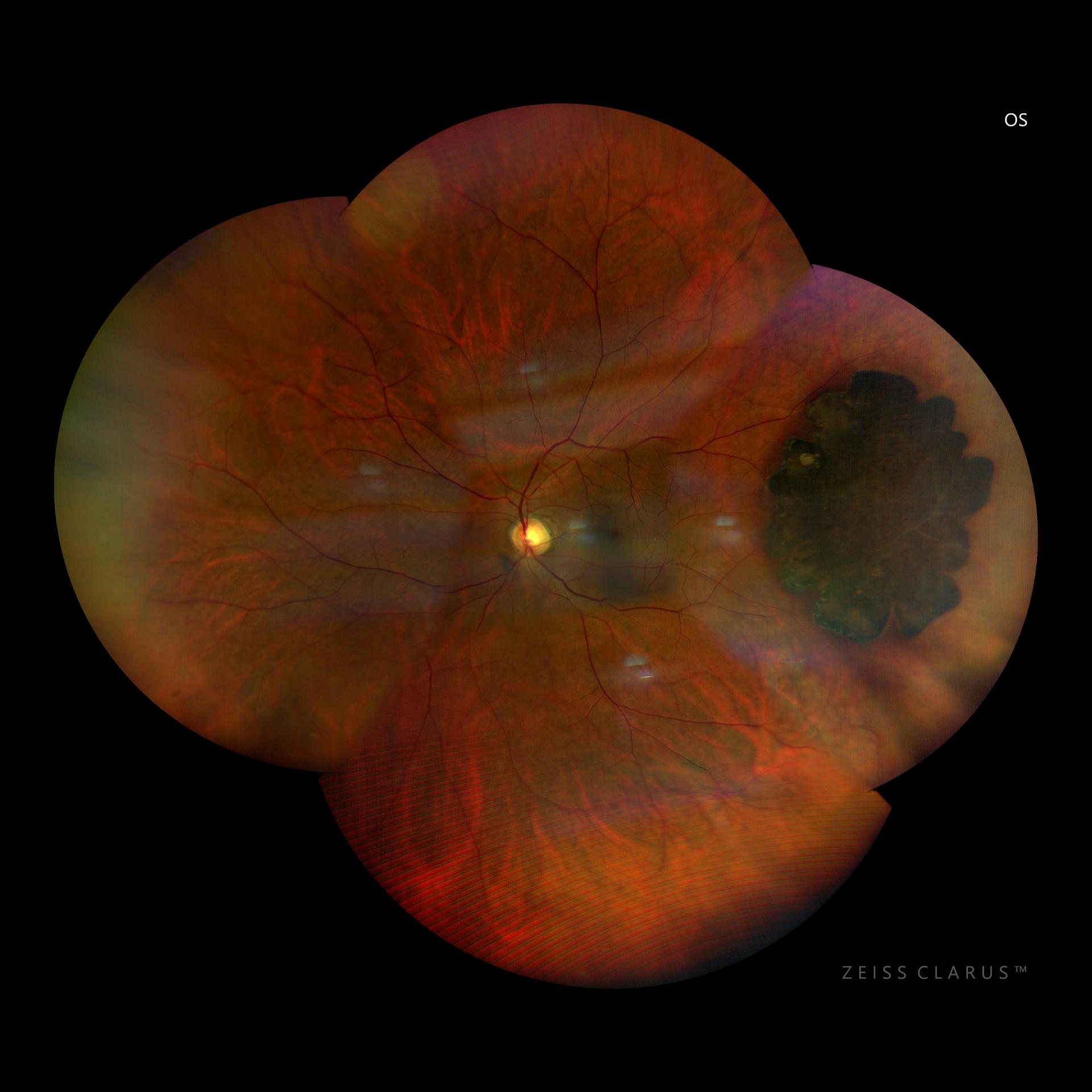
Case of IOL Dislocation
Retinal analysis of a 65-year-old male was diagnosed with dislocated IOL. The auto-montage imaging revealed 4 haptic akreos IOL along with capsular bag lying flat in the inferior vitreous cavity. The patient had a prior history of vitrectomy for RD.
Case of Subhyaloid Hemorrhage
Retinal analysis (true color) of a 24-year-old female diagnosed with Subhyaloid Hemorrhage post Valsalva. The Wide Field fundus image (figure 1) allowed the identification of sub hyaloid hemorrhage located at macula. Treatment was performed by YAG hyaloidotomy. Post laser WF imaging (figure 2) shows drainage of blood into the inferior vitreous cavity producing immediate resolution.
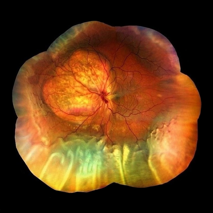
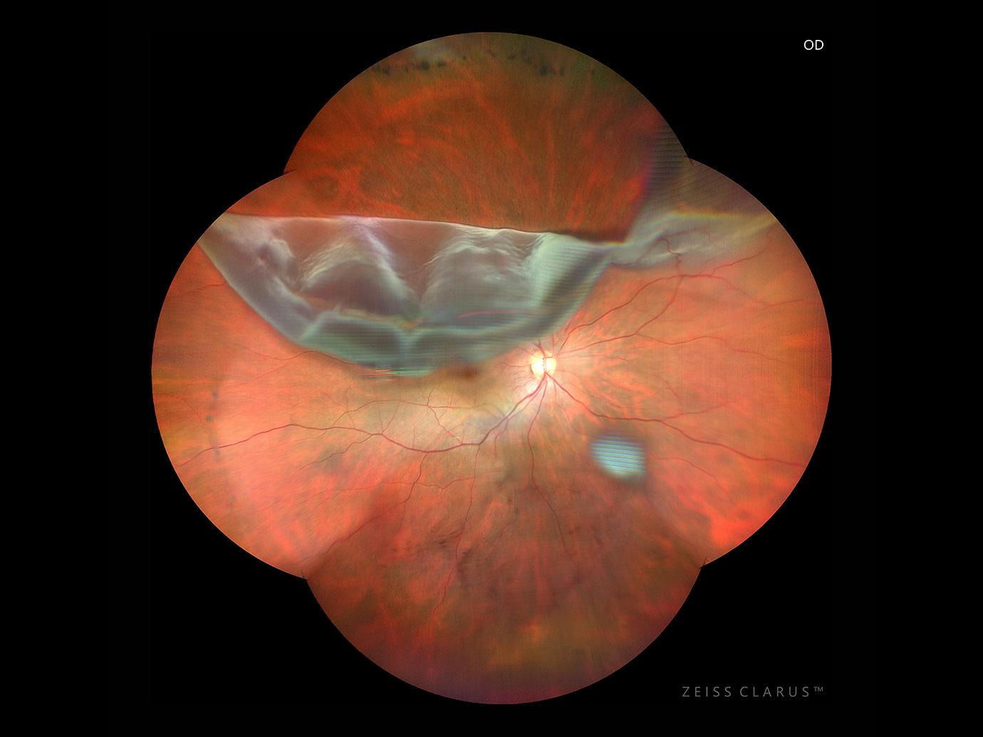
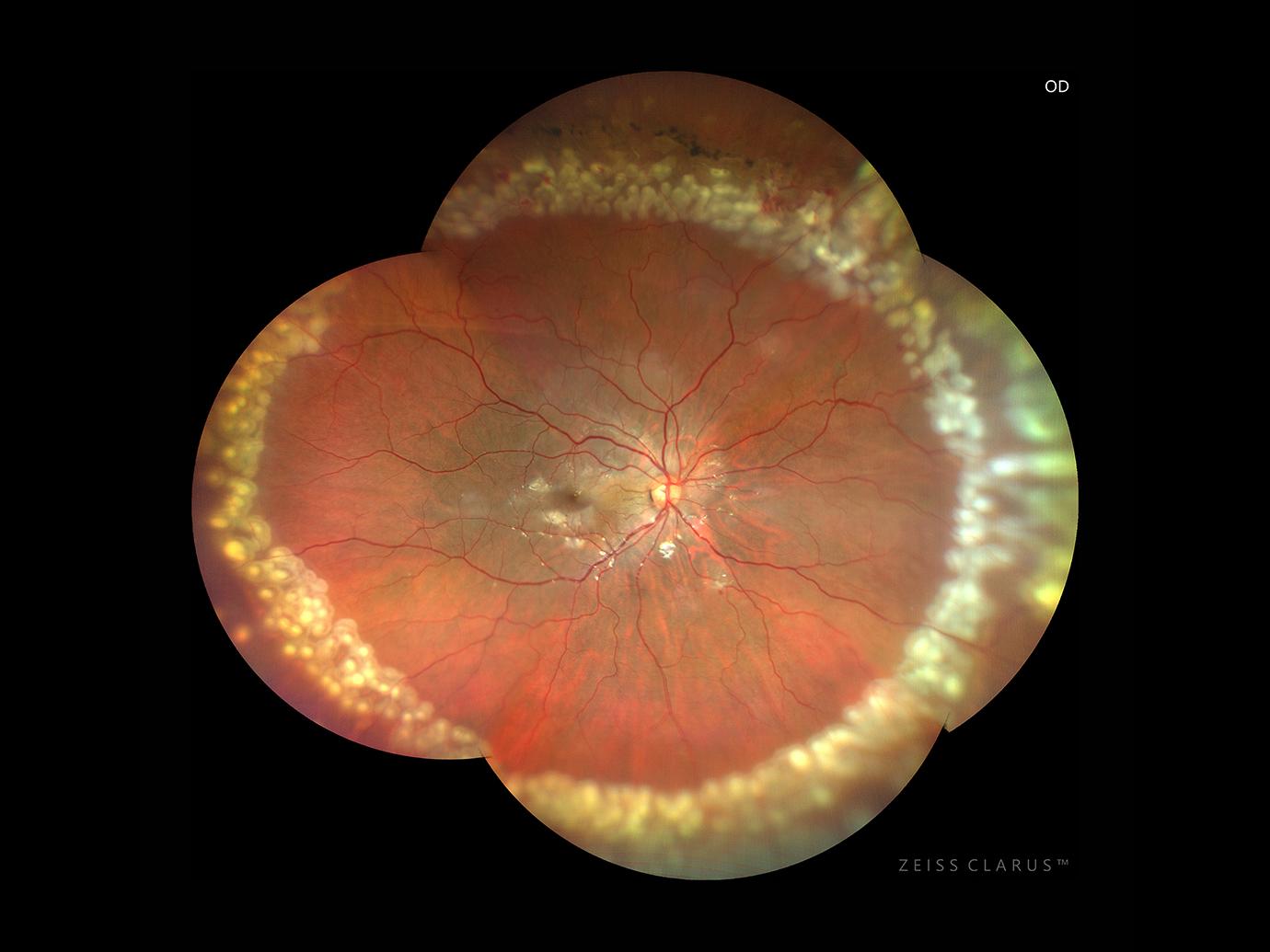
Case of Rhegmatogenous Retinal Detachment
Retinal analysis of a 56-year-old male diagnosed with Rhegmatogenous Retinal Detachment. True color auto-montage image (figure 1) shows a retinal detachment with superior giant retinal tear. Treatment was performed by vitreoretinal surgery under ophthalmic surgical liquid. Post-surgery auto-montage image (figure 2) shows attached retina and fresh laser marks.
When I explored the capabilities of ZEISS CLARUS, I was impressed by the picture quality, from the macula to the periphery. Incorporating ZEISS CLARUS into my practice streamlined diagnostics and provided insights into retinal and optic nerve conditions that were previously difficult to assess. Acquiring ZEISS CLARUS aligns with my goal of providing the best care to my patients.
Third-party Content Blocked
The video player is blocked due to your cookie preferences. To change the settings and play the video, please click the button below and consent to use of "Functional" tracking technologies.
Discover how ZEISS CLARUS has revolutionized Dr. Vishal's approach to retina care
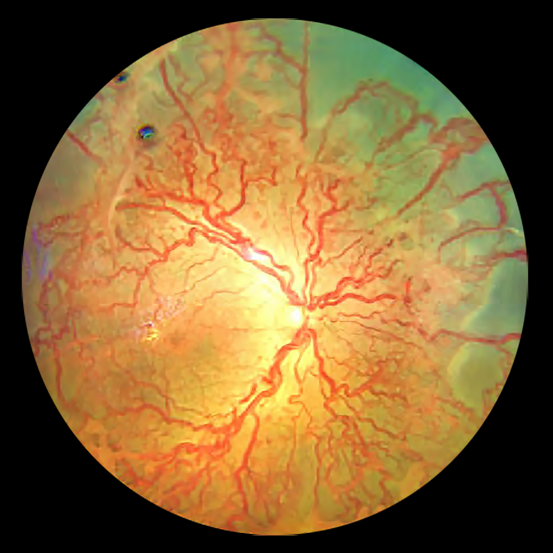
Non-contact wide-field fundus imaging for ROP
Imaging the retina is important in Retinopathy of Prematurity (ROP) not only for documentation, follow-up, teleconsultation, sharing with parents and for teaching students, but also from medico-legal perspectives.
Fundus photography is all about the 'color'. While a high resolution and widefield of view are a pre-requisite to get all the information, the true colors are an important component for accurate interpretation and diagnosis. Most of the dedicated neonatal fundus cameras are 'contact' systems and the 'pseudo-colors' in some cameras can camouflage the true nature of the disease.
The ZEISS CLARUS 700 which uses true color reflectance imaging with high resolution and a non-contact approach, hence has a great potential for fundus photography of newborn babies in addition to its current usefulness in adult retinal diseases.
Third-party Content Blocked
The video player is blocked due to your cookie preferences. To change the settings and play the video, please click the button below and consent to use of "Functional" tracking technologies.
Unedited Real time image capture by ZEISS CLARUS 700
ROP imaging technique with ZEISS CLARUS
The ZEISS CLARUS 700 imaging system lends itself readily to neonatal retinal imaging. The fast rate of image acquisition and non-contact nature are comfortable and safe for the baby. The ZEISS CLARUS 700 which uses true color reflectance imaging with high resolution and a non-contact approach, hence has a great potential for fundus photography of newborn babies in addition to its current usefulness in adult retinal diseases.
A glimpse into the world of ROP - LVPEI Atlas
Anant Bajaj Retina Institute diagnostic team at LVPEI under the leadership of Dr. Akash Belenje and Mr. R Ugandhar Reddy have demonstrated in this atlas something unique and is published for the very first time. The atlas contains 100 ROP images of varying disease severity and in different clinical situations. The true color reflectance images with high resolution provide a wonderful opportunity to see the entire spectrum of ROP presentations, follow-up, pre and post treatment and its natural history.
CLARUS 700 from ZEISS was designed as a comprehensive ultra-widefield retinal camera for eye care specialists to capture ultra-widefield images in true color, with unsurpassed image quality and a complete suite of modalities including fluorescein angiography.